CESSYS® – Cervical Endoscopic Surgical System
The endoscopic system for the treatment of disc herniations and spinal stenosis of the cervical spine.
The CESSYS® Family
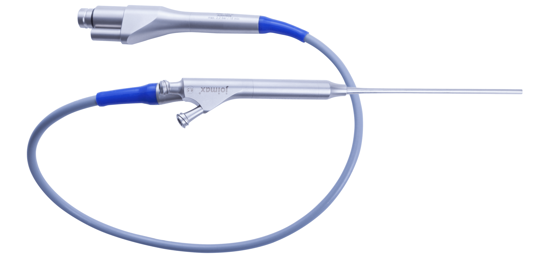
The CESSYS® family offers a comprehensive solution for deherniation and decompression of the cervical spine.
The combination of CESSYS® dorsal, posterior access, and CESSYS® ventral, anterior access, provides an almost 360° access of the cervical spinal canal.
The CESSYS® Family
The CESSYS® family offers a comprehensive solution for deherniation and decompression of the cervical spine.
The combination of CESSYS® dorsal, posterior access, and CESSYS® ventral, anterior access, provides an almost 360° access of the cervical spinal canal.

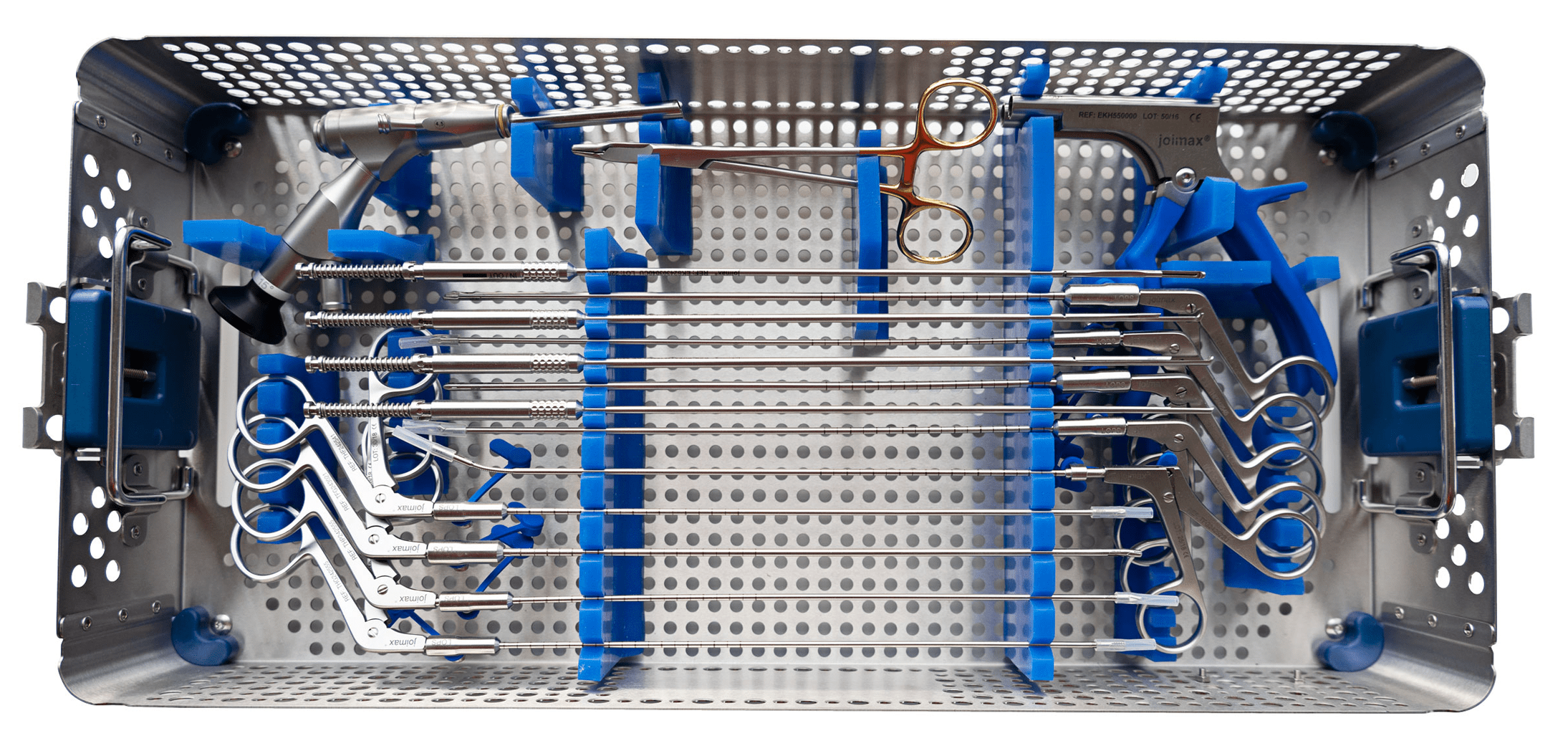
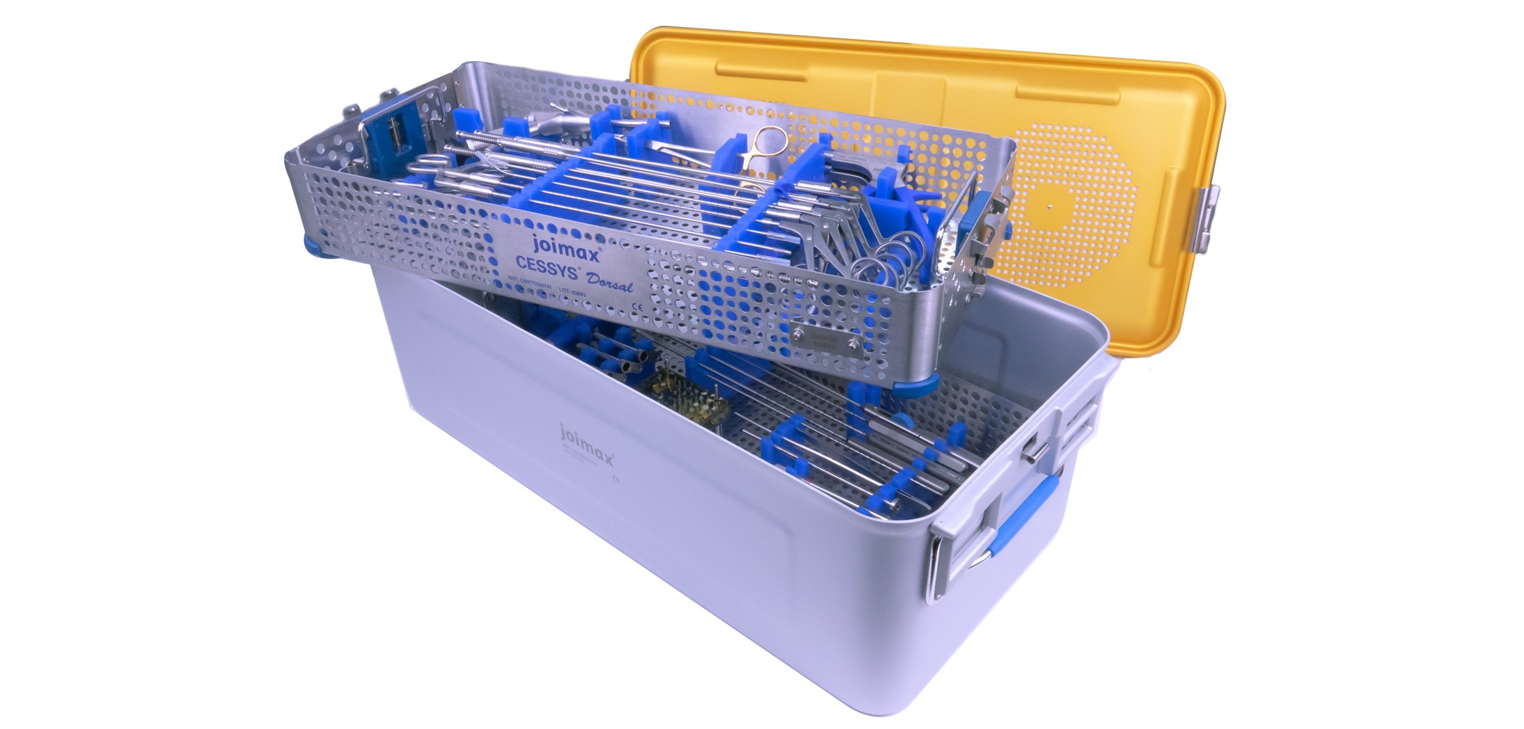
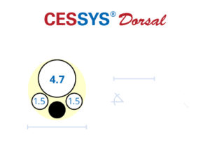
The CESSYS® Dorsal instrument set contains all reusable instruments for the posterior access to the spinal canal, the palpation and preparation of tissue structures as well as the resection of the pathology.
Grasping, cutting, and punching forceps facilitate the removal of soft tissue while special Endo-Kerrisons punches offer the necessary bite for the removal of bone structures.

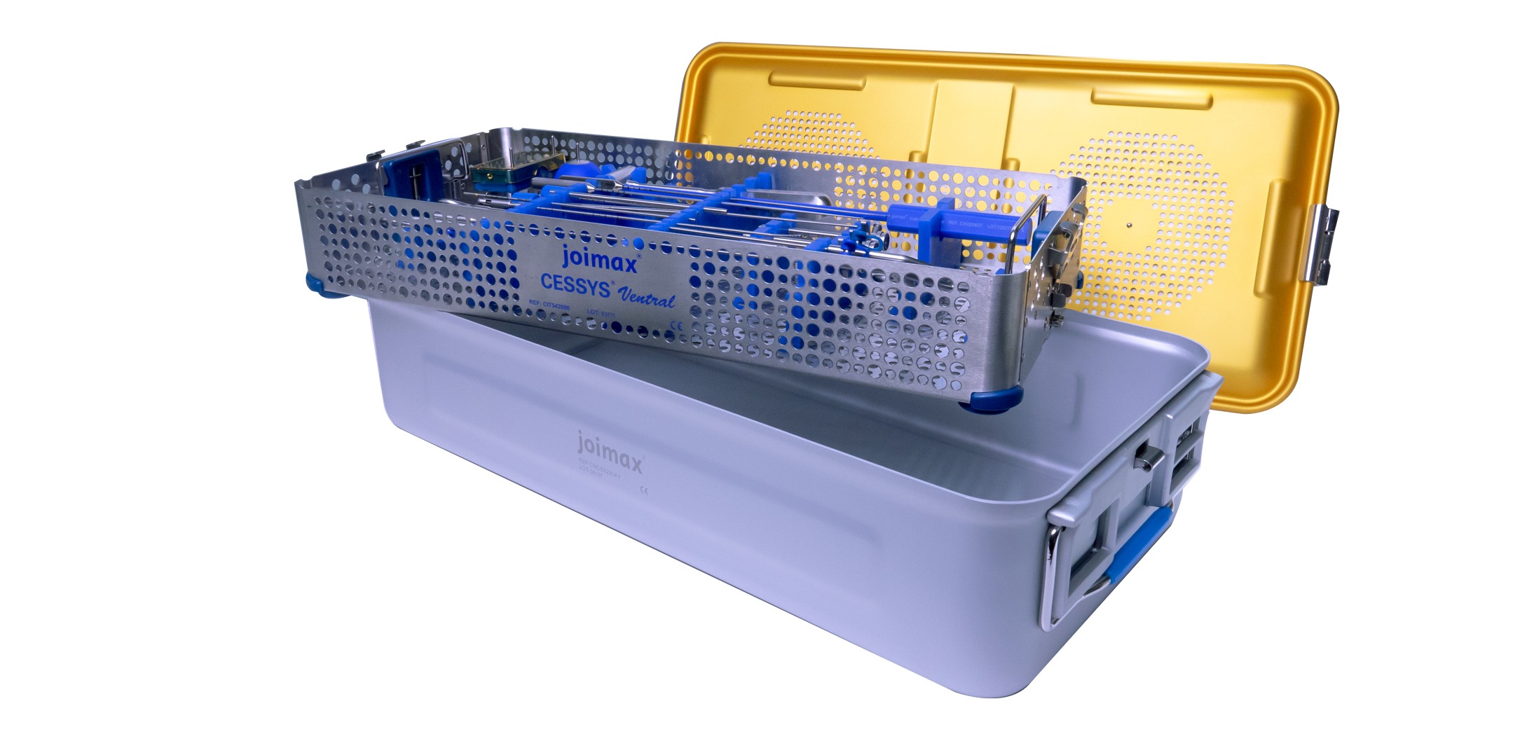
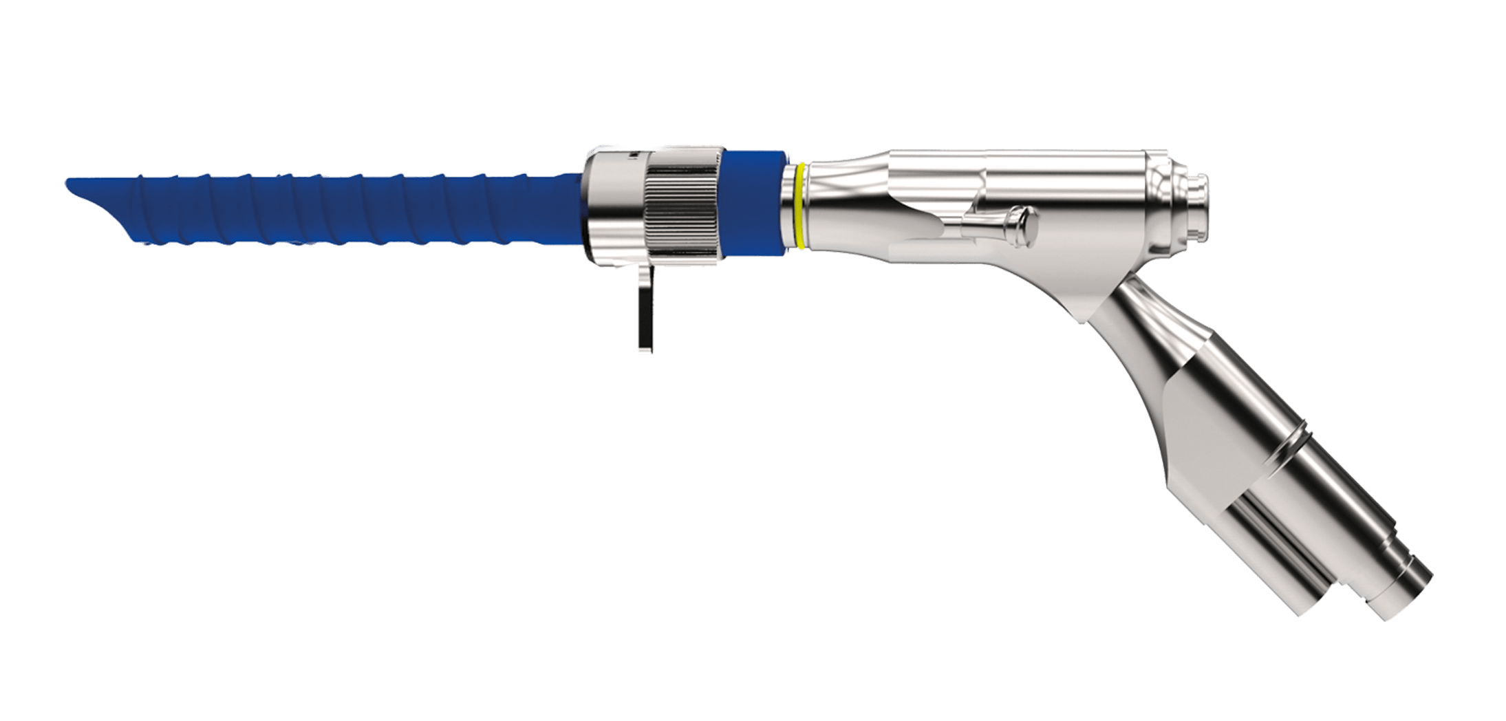
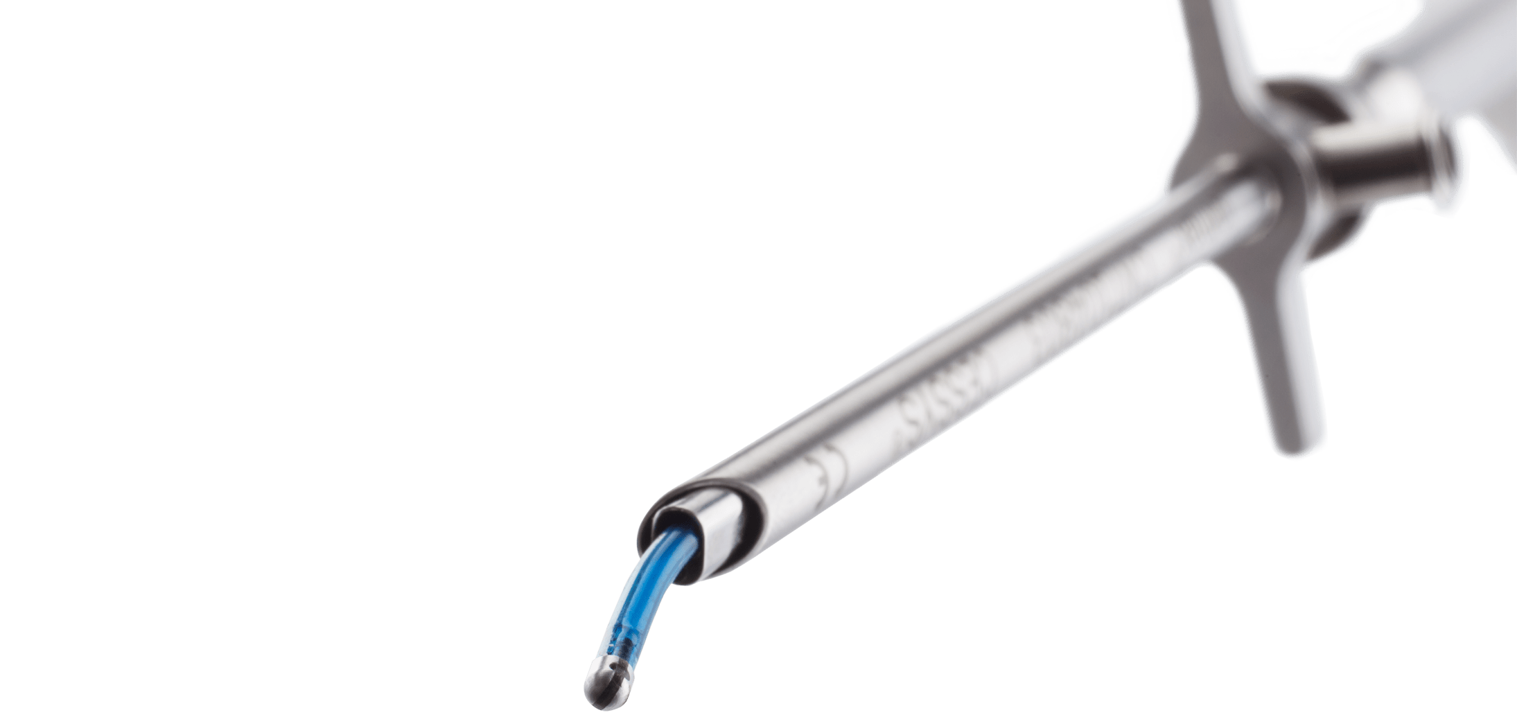
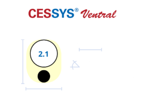
The CESSYS® Ventral instrument set is specifically designed for the anterior access of the cervical spine.
The set is available in two variants with either round working tubes for high mobility or oval working tubes with minimized height for tighter intradiscal spaces.
The CESSYS® Surgery
CESSYS® Dorsal – Posterior Access
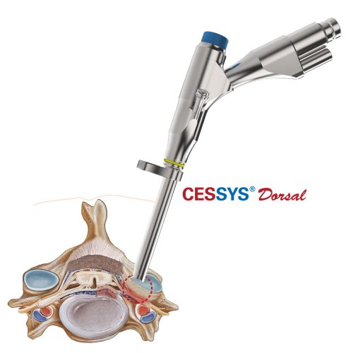
The CESSYS® Dorsal posterior approach, similar to the iLESSYS® interlaminar technique, utilizes the interlaminar window for access.
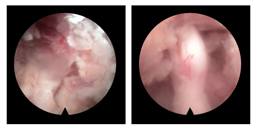
It provides a safe access route through which an endoscope can directly access pathologies within the spinal canal.

Selected Indications
• Herniated intervertebral disc (protrusion, prolapse, sequester and free fragment)
• Spinal stenosis through soft and/or bone tissue
• Facet joint cyst

Selected Indications
• Herniated intervertebral disc (protrusion, prolapse, sequester and free fragment)
• Spinal stenosis through soft and/or bone tissue
• Facet joint cyst

CESSYS® Ventral – Anterior Access
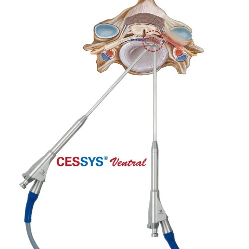
The CESSYS® Ventral method uses an anterior approach through the disc space directly at the site of the herniation.
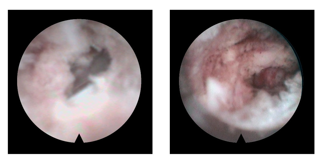
Bulging, compressing disc material can be removed endoscopically under direct visual control without the need for a laminotomy. CESSYS® Ventral is the perfect tool for soft lateral and mediolateral herniations as well as
mild ventral stenosis.
CESSYS® Ventral – Anterior Access
The CESSYS® Ventral method uses an anterior approach through the disc space directly at the site of the herniation.
Bulging, compressing disc material can be removed endoscopically under direct visual control without the need for a laminotomy. CESSYS® Ventral is the perfect tool for soft lateral and mediolateral herniations as well as mild ventral stenosis.

Selected Indications
• Herniated intervertebral disc (protrusion und prolapse, sequester)
• Mild spinal stenosis through soft and/or bone tissue
Selected Indications
• Herniated intervertebral disc (protrusion und prolapse, sequester)
• Mild spinal stenosis through soft and/or bone tissue

Selected Indications
• Herniated intervertebral disc (protrusion und prolapse, sequester)
• Mild spinal stenosis through soft and/or bone tissue
Scientific Literature for CESSYS®

[1] Learning curve for endoscopic posterior cervical foraminotomy
Perfetti DC, Rogers-LaVanne MP, Satin AM, Yap N, Khan I, Kim P, Hofstetter CP, Derman PB; Eur Spine J (2023): doi:10.1007/s00586-023-07623-6
[2] Microscopic Anterior Cervical Discectomy and Fusion Versus Posterior Percutaneous Endoscopic Cervical Keyhole Foraminotomy for Single-level Unilateral Cervical Radiculopathy: A Systematic Review and Meta-analysis
Guo L, Wang J, Zhao Z, Li J, Zhao H, Gao Y, Chen C; Clinical Spine Surgery (2023) 36: 59
[3] The first experience with fully endoscopic posterior cervical foraminotomy and discectomy for radiculopathy performed in Viet Duc University Hospital
Dinh SN, Dinh HT; Sci Rep (2022) 12: 8314
[4] Clinical efficacy and learning curve of posterior percutaneous endoscopic cervical laminoforaminotomy for patients with cervical spondylotic radiculopathy
Yao R, Yan M, Liang Q, Wang H, Liu Z, Li F, Zhang H, Li K, Sun F; Medicine (2022) 101:e30401
[5] Comparison of Percutaneous Endoscopic Cervical Keyhole Foraminotomy versus Microscopic Anterior Cervical Discectomy and Fusion for Single Level Unilateral Cervical Radiculopathy
Ma W, Peng Y, Zhang S, Wang Y, Gan K, Zhao X, Xu D; IJGM (2022) 15: 6897–6907
[6] Clinical Efficacy of Posterior Percutaneous Endoscopic Unilateral Laminotomy with Bilateral Decompression for Symptomatic Cervical Spondylotic Myelopathy
Zhao X-B, Ma Y-J, Ma H-J, Zhang X-Y, Zhou H-G; Orthop Surg (2022): doi:10.1111/
os.13237
[7] Outcome of Anterior and Posterior Endoscopic Procedures for Cervical Radiculopathy Due to Degenerative Disk Disease: A Systematic Review and Meta-Analysis
Alomar SA, Maghrabi Y, Baeesa SS, Alves ÓL; Global Spine J (2021); 21925682211037270
[8] Comparative evaluation of posterior percutaneous endoscopy cervical discectomy using a 3.7 mm endoscope and a 6.9 mm endoscope for cervical disc herniation: a retrospective comparative cohort study
Yu T, Wu J-P, Zhang J, Yu H-C, Liu Q-Y; BMC Musculoskelet Disord (2021) 22: 131
[9] Full endoscopic cervical spine surgery
Shen J, Telfeian AE, Shaaya E, Oyelese A, Fridley J, Gokaslan ZL; J Spine Surgery (2020) 6(2): 383–390
[10] Full endoscopic unilateral laminotomy for bilateral decompression of the cervical spine: surgical technique and early experience
Carr DA, Abecassis IJ, Hofstetter CP; J Spine Surg (2020) 6: 447–456
- Ma W, Peng Y, Zhang S, Wang Y, Gan K, Zhao X et al. Comparison of Percutaneous Endoscopic Cervical Keyhole Foraminotomy versus Microscopic Anterior Cervical Discectomy and Fusion for Single Level Unilateral Cervical Radiculopathy. IJGM 2022; 15: 6897–6907.
- Dinh SN, Dinh HT. The first experience with fully endoscopic posterior cervical foraminotomy and discectomy for radiculopathy performed in Viet Duc University Hospital. Sci Rep 2022; 12: 8314.
- Shen J, Shaaya E, Bae J, Telfeian AE. Endoscopic Spine Surgery of the Cervicothoracic Spine: A Review of Current Applications. Int J Spine Surg 2021; 15: S93–S103.
- Brown GSC, Gibson JNA. Early experience with cervical endoscopic spinal surgery (CESS): a potential adjunct to ACDF/disc arthroplasty. The Spine Journal 2015; 15: S74.
- Liu Y, Tang G-K, Wang W-H, Shi C-G, Wang S, Yu L et al. Morphology of Herniated Disc as a Predictor for Outcomes of Posterior Percutaneous Full-endoscopic Cervical Discectomy in Treating Cervical Spondylotic Radiculopathy. Orthop Surg 2021. https://doi.org/10.1111/os.13134.
- Hofstetter CP. Atlas of Full-Endoscopic Spine Surgery. 2020.https://www.thieme.com/books-main/neurosurgery/product/5466-atlas-of-full-endoscopic-spine-surgery (accessed 6 Aug2021).
- Middleton SD, Wagner R, Gibson JA. Multi-level spine endoscopy: a review of available evidence and case report. EFORT open reviews 2017; 2: 317–323.
- Shen J, Telfeian AE, Shaaya E, Oyelese A, Fridley J, Gokaslan ZL. Full endoscopic cervical spine surgery. J Spine Surg 2020; 6: 383–390.
